

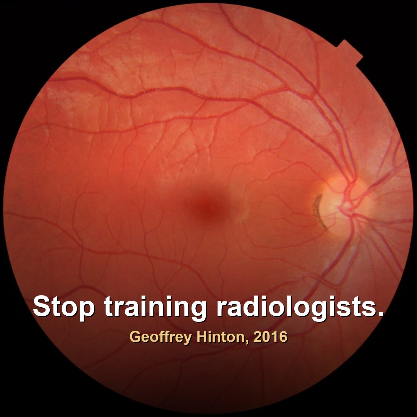
Ten years ago, Geoffrey Hinton, now a Nobel laureate in Physics and one of the architects of modern AI, gave the field a famous warning: stop training radiologists, because deep learning would out-read them within five years. He has since refined that view, and history has been kinder to radiologists than to his timing. A decade on, demand for them is growing faster than almost any profession.
So why did such a sharp prediction miss? The clearest place to look is the eye, where AI already reads scans better than we can. And yet the eye-care workforce is still growing too. What happens to the next generation of doctors who never get to read the “easy” cases?
The Curioso in me went deep on this over the past week. Here is what I learned, and the people and institutions worth following.
The numbers have moved fast
A friend of mine worked on one of the early AI ophthalmology engines. Around 2018, after two to three years and millions of scans, it crossed 80% accuracy, already ahead of the human ophthalmologists in their sample. Today, FDA-cleared screening tools average roughly 93% sensitivity and 90 to 93% specificity across very large real-world datasets, and some systems push sensitivity past 95% while staying deliberately conservative on safety. The machines rarely miss the serious stuff. (New to terms like sensitivity, specificity or FDA-cleared? A plain-English glossary sits at the end.)
The counterintuitive part
Despite all that automation, ophthalmologist and radiologist jobs have not fallen. AI has absorbed the high-volume, routine screening, especially for diabetic retinopathy, and handed specialists back their time for treatment decisions, injections, surgery and complex cases. One randomised trial found AI screening let a clinic complete around 40% more eye exams per hour. Demand keeps climbing because of rising diabetes prevalence and an ageing population.
But here is the honest version. If you normalise for that demand, hold population and disease rates flat, the headcount effect of pure automation becomes visible, and it is modestly negative. Fewer full-time specialists would be needed for screening, and new graduates would face tougher competition. We should not hide behind the demand curve when we talk about the long run.
The real worry is training, not replacement
If AI reads most of the routine “bread and butter” cases, how do junior doctors build the pattern recognition that turns them into experts? This deskilling risk is real and openly debated. The best programmes are responding by pushing trainees earlier into complex cases, interventional work and multidisciplinary boards, and by teaching residents to build and test AI themselves, so they shape the technology instead of inheriting it.
Who is doing the best thinking
United States. Emory Dept of Radiology runs the strongest dedicated model, a four-year integrated imaging-informatics track woven through residency. UT Southwestern Radiology teaches AI to all its residents and runs an advanced track for those who want depth. Nationally, RSNA and the ACR Data Science Institute (part of American College of Radiology) set much of the curriculum standard.
United Kingdom. King’s College London, through its AI Centre for Value Based Healthcare, pairs with the Royal College of Radiologists on hands-on training. The National Imaging Academy Wales is embedding AI into structured pathways.
Three UK voices I would follow on exactly this question:
· Gerald Lip frames AI as a safety net in breast screening: it cuts the routine workload, while humans stay essential for oversight and for catching model drift.
· Susan Shelmerdine flags the paediatric blind spot: most tools are trained on adults and perform poorly on children, so she pushes for child-specific datasets and ethical deployment.
· Thomas Booth, AI Faculty Lead for the Royal College of Radiologists, is the practical educator: radiologists need real technical training to build and evaluate AI, not just use it.
A deliberate detour to Manila, and a personal one. I have lived in eight countries and worked in around fifty, and the part of this story I care most about is the global south, the low- and middle-income countries where vast populations are underserved and a single screening tool can matter more than anywhere else. Diabetic retinopathy blinds people who never get near an ophthalmologist. If autonomous AI earns its keep anywhere, it has to earn it there. I spend a good part of each year in the Philippines, mentoring founders and operators, so I watch this market closely.
Philippines (Manila). The Philippine College of Radiology leads nationally, having published AI guidelines and stood up a dedicated AI subcommittee, and The Medical City is among the hospitals bringing AI imaging tools into local practice. Three Manila professionals worth knowing:
· Erwin John T. Carpio sits on the PCR AI subcommittee and co-drafted the College’s AI guidelines.
· Ruben G. Kasala, an endocrinologist and CEO of The Medical City Ortigas, represents the hospital-leadership side of AI adoption.
· Raymond Francis R. Sarmiento, Director of the UP Manila National Telehealth Center, writes on responsible AI adoption and health policy.
A spotlight on a friend
The first person I ever spoke to about this, back around 2017 when the Curioso in me went to visit him at the hospital, was Pearse Keane. He is now Professor of Artificial Medical Intelligence at the UCL Institute of Ophthalmology, a consultant ophthalmologist at Moorfields Eye Hospital, and Director and founder of the INSIGHT Health Data Research Hub, the world’s largest ophthalmology bioresource. In 2025 he was elected to the US National Academy of Medicine and awarded The Royal Society‘s Gabor Medal for his work in AI ophthalmology and oculomics, using the eye to detect systemic disease elsewhere in the body. In March he spoke at 100% Optical in London on transforming ophthalmology with AI, and keynoted EURETINA‘s Special Focus Meeting on AI in Retina, also in London. This October he co-chairs the AI session at EURETINA’s Congress in Vienna. His team is now scaling a national oculomics project, backed by 3.7 million pounds of MRC and NIHR funding to link eye-scan data across the NHS. An original mind, and a generous one.
Efficiency now, expertise later
There is a quiet conflict between short-term efficiency and long-term workforce planning. The optimists, and I lean that way, believe the clinicians who master AI will become more valuable, not less, because they will shape the tools and know when to override them. But that only holds if we protect the training pipeline now, before a generation grows up never having read the cases that build judgement.
Hinton, to his credit, has kept thinking out loud. Last year he warned that for mundane intellectual labour ‘AI is just going to replace everybody’, and that the safest trade now is plumbing. On radiology he may prove to have been early rather than wrong, and that possibility deserves respect.
If you work in radiology, ophthalmology or medical AI, I would love your view. Are we protecting the next generation’s path to expertise, or quietly automating it away?
Coming next: in a future post the Curioso in me will turn this same lens on other careers, the places where AI’s effect is already showing up at scale, sometimes brutally, sometimes in ways nobody predicted. Language interpreters, paralegals, GPs, professional drivers, teachers. If you have hard evidence of real-world impact in any of those fields, positive or negative, send it my way. It will feed the deep dives.
Glossary (plain English, for the curious)
Sensitivity. how often a test correctly catches the people who DO have the disease. High sensitivity means few missed cases.
Specificity. how often a test correctly clears the people who do NOT have the disease. High specificity means few false alarms.
Diabetic retinopathy. damage to the retina (the light-sensitive layer at the back of the eye) caused by diabetes. A leading cause of preventable blindness, and the main thing these AI tools screen for.
Retinal scan (fundus image). a photograph of the inside back of the eye, where the retina, its blood vessels and the optic nerve are visible.
Oculomics. reading the eye to detect disease elsewhere in the body, for example heart disease or dementia, because the eye’s vessels and nerves are a window onto the rest of you.
Bioresource. a large, curated library of medical data, here millions of eye images, gathered so researchers can train and test tools on it.
FDA-cleared. formally authorised by the US medicines and devices regulator for use in real clinical practice.
Autonomous AI screening. AI that makes the screening call itself, without a clinician needing to read every single scan.
Deskilling. the gradual loss of a skill when people stop practising it, the core worry for trainees if AI reads all the routine cases.
Links referenced
People
Institutions
· UCL Institute of Ophthalmology
· INSIGHT Health Data Research Hub
· US National Academy of Medicine
· The Royal Society (Gabor Medal)
· EURETINA
· Emory Department of Radiology
· RSNA
· AI Centre for Value Based Healthcare
· Royal College of Radiologists
· National Imaging Academy Wales
Source